Welcome to the Sai Lab!
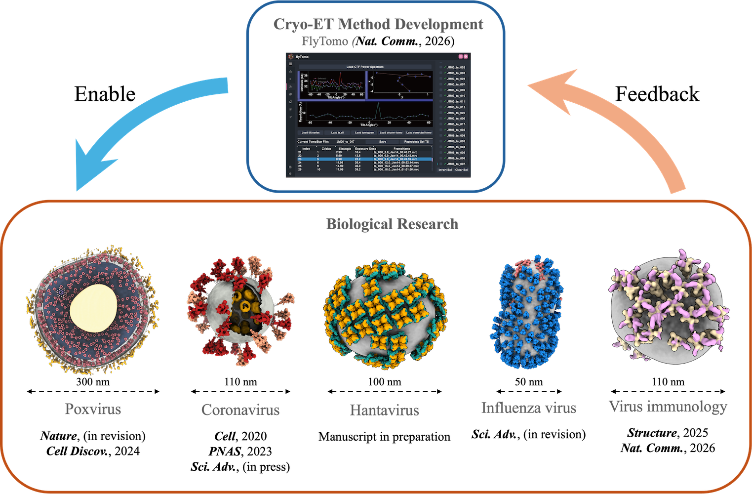
Understanding biological macromolecules requires investigating their native cellular contexts. Cryo-electron tomography (cryo-ET) is a transformative in-situ technique, yet its broad application is limited by low resolution and throughput. Meanwhile, enveloped viruses (e.g., SARS-CoV-2, influenza, Poxvirus) present complex, flexible architectures that remain challenging for traditional structural methods.
Our lab integrates cryo-ET method development with the in-situ structural biology of enveloped viruses. By overcoming technical bottlenecks in cryo-ET, we provide new structural insights for fundamental virology and therapeutic applications.
1. High-Resolution Cryo-ET Methodology
We advance high-resolution cryo-ET and subtomogram averaging. Combined with CLEM and cryo-FIB, our computational and experimental workflows bridge structural biology across scales—from intact virions to cellular environments.
2. In-Situ Architecture & Function of Emerging Viruses
Using cryo-ET, we systematically dissect the in-situ mechanisms of viral assembly, infection, and immune recognition. We aim to expose viral vulnerabilities to guide rational antibody and vaccine design.
🔥 HIGHLIGHT · LATEST PUBLICATION
FlyTomo: a streamlined software for on-the-fly cryo-ET data processing and diagnosis
Nature Communications, 2026
✨ Key findings:
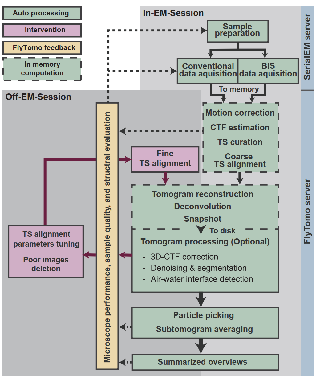
Cryo-ET combined with subtomogram averaging (STA) enables the structural elucidation of macromolecular assemblies in native environments. However, their widespread adoption has been limited by the labor-intensive, expertise-dependent data processing workflow. Here we present FlyTomo, a software that streamlines data processing from frame alignment to STA with high-throughput for authentic cryo-ET scenarios. During data acquisition, FlyTomo performs real-time diagnosis, enabling prompt feedback on sample quality, microscope performance and structural features. After acquisition, it aggregates diagnostic metrics into an overview, guiding users through data review and refinement. FlyTomo also curates raw and processed data into directories to simplify data management and archiving. We validate FlyTomo across a diverse set of authentic cryo-ET samples, including purified enveloped viruses and cryo-lamellae, on multiple microscopes and cameras, achieving structures at resolutions of 3.4 to 7.3 Å. Collectively, by integrating accuracy, scalability and usability, FlyTomo reduces the technical barrier for in situ structural biology using cryo-ET.